What Structure Allows Air to and From the Lungs?
The Respiratory System
The Lungs
Learning Objectives
By the end of this section, you lot volition be able to:
- Describe the overall function of the lung
- Summarize the blood menstruation pattern associated with the lungs
- Outline the beefcake of the claret supply to the lungs
- Draw the pleura of the lungs and their office
A major organ of the respiratory organisation, each lung houses structures of both the conducting and respiratory zones. The chief role of the lungs is to perform the exchange of oxygen and carbon dioxide with air from the atmosphere. To this finish, the lungs exchange respiratory gases across a very large epithelial surface expanse—near 70 square meters—that is highly permeable to gases.
Gross Anatomy of the Lungs
The lungs are pyramid-shaped, paired organs that are continued to the trachea by the right and left bronchi; on the inferior surface, the lungs are bordered by the diaphragm. The diaphragm is the flat, dome-shaped musculus located at the base of the lungs and thoracic crenel. The lungs are enclosed by the pleurae, which are attached to the mediastinum. The right lung is shorter and wider than the left lung, and the left lung occupies a smaller volume than the right. The cardiac notch is an indentation on the surface of the left lung, and it allows space for the middle ((Figure)). The noon of the lung is the superior region, whereas the base is the reverse region almost the diaphragm. The costal surface of the lung borders the ribs. The mediastinal surface faces the midline.
Gross Anatomy of the Lungs

Each lung is composed of smaller units called lobes. Fissures separate these lobes from each other. The right lung consists of iii lobes: the superior, middle, and inferior lobes. The left lung consists of two lobes: the superior and inferior lobes. A bronchopulmonary segment is a sectionalisation of a lobe, and each lobe houses multiple bronchopulmonary segments. Each segment receives air from its ain tertiary bronchus and is supplied with blood by its own artery. Some diseases of the lungs typically touch one or more bronchopulmonary segments, and in some cases, the diseased segments tin can be surgically removed with little influence on neighboring segments. A pulmonary lobule is a subdivision formed as the bronchi branch into bronchioles. Each lobule receives its own large bronchiole that has multiple branches. An interlobular septum is a wall, composed of connective tissue, which separates lobules from one another.
Blood Supply and Nervous Innervation of the Lungs
The blood supply of the lungs plays an of import role in gas substitution and serves as a transport system for gases throughout the body. In addition, innervation by the both the parasympathetic and sympathetic nervous systems provides an of import level of control through dilation and constriction of the airway.
Blood Supply
The major role of the lungs is to perform gas exchange, which requires claret from the pulmonary circulation. This blood supply contains deoxygenated blood and travels to the lungs where erythrocytes, also known as red claret cells, pick up oxygen to be transported to tissues throughout the body. The pulmonary artery is an avenue that arises from the pulmonary body and carries deoxygenated, arterial blood to the alveoli. The pulmonary avenue branches multiple times every bit it follows the bronchi, and each co-operative becomes progressively smaller in diameter. 1 arteriole and an accompanying venule supply and drain one pulmonary lobule. As they near the alveoli, the pulmonary arteries become the pulmonary capillary network. The pulmonary capillary network consists of tiny vessels with very thin walls that lack smooth muscle fibers. The capillaries branch and follow the bronchioles and structure of the alveoli. It is at this point that the capillary wall meets the alveolar wall, creating the respiratory membrane. Once the blood is oxygenated, it drains from the alveoli by way of multiple pulmonary veins, which exit the lungs through the hilum.
Nervous Innervation
Dilation and constriction of the airway are accomplished through nervous command past the parasympathetic and sympathetic nervous systems. The parasympathetic arrangement causes bronchoconstriction, whereas the sympathetic nervous system stimulates bronchodilation. Reflexes such as coughing, and the power of the lungs to regulate oxygen and carbon dioxide levels, also result from this autonomic nervous system control. Sensory nervus fibers arise from the vagus nerve, and from the 2nd to fifth thoracic ganglia. The pulmonary plexus is a region on the lung root formed by the archway of the nerves at the hilum. The fretfulness then follow the bronchi in the lungs and branch to innervate muscle fibers, glands, and blood vessels.
Pleura of the Lungs
Each lung is enclosed within a crenel that is surrounded by the pleura. The pleura (plural = pleurae) is a serous membrane that surrounds the lung. The right and left pleurae, which enclose the right and left lungs, respectively, are separated by the mediastinum. The pleurae consist of two layers. The visceral pleura is the layer that is superficial to the lungs, and extends into and lines the lung fissures ((Figure)). In contrast, the parietal pleura is the outer layer that connects to the thoracic wall, the mediastinum, and the diaphragm. The visceral and parietal pleurae connect to each other at the hilum. The pleural cavity is the space betwixt the visceral and parietal layers.
Parietal and Visceral Pleurae of the Lungs
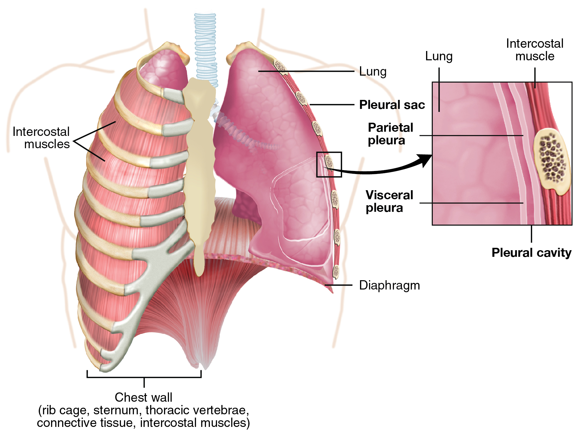
The pleurae perform two major functions: They produce pleural fluid and create cavities that carve up the major organs. Pleural fluid is secreted by mesothelial cells from both pleural layers and acts to lubricate their surfaces. This lubrication reduces friction between the two layers to prevent trauma during animate, and creates surface tension that helps maintain the position of the lungs against the thoracic wall. This adhesive characteristic of the pleural fluid causes the lungs to enlarge when the thoracic wall expands during ventilation, allowing the lungs to fill up with air. The pleurae also create a sectionalization between major organs that prevents interference due to the move of the organs, while preventing the spread of infection.
Everyday Connection
The Effects of Second-Hand Tobacco Smoke The burning of a tobacco cigarette creates multiple chemical compounds that are released through mainstream smoke, which is inhaled past the smoker, and through sidestream smoke, which is the fume that is given off by the burning cigarette. Second-hand smoke, which is a combination of sidestream fume and the mainstream smoke that is exhaled by the smoker, has been demonstrated by numerous scientific studies to cause disease. At least 40 chemicals in sidestream smoke have been identified that negatively impact human health, leading to the development of cancer or other atmospheric condition, such as allowed system dysfunction, liver toxicity, cardiac arrhythmias, pulmonary edema, and neurological dysfunction. Furthermore, 2d-paw smoke has been constitute to harbor at to the lowest degree 250 compounds that are known to be toxic, carcinogenic, or both. Some major classes of carcinogens in second-manus smoke are polyaromatic hydrocarbons (PAHs), N-nitrosamines, effluvious amines, formaldehyde, and acetaldehyde.
Tobacco and second-manus smoke are considered to be carcinogenic. Exposure to second-hand smoke tin can cause lung cancer in individuals who are not tobacco users themselves. It is estimated that the take a chance of developing lung cancer is increased by upward to 30 pct in nonsmokers who live with an private who smokes in the house, as compared to nonsmokers who are non regularly exposed to second-mitt smoke. Children are especially affected past second-mitt smoke. Children who live with an individual who smokes inside the domicile take a larger number of lower respiratory infections, which are associated with hospitalizations, and higher risk of sudden infant expiry syndrome (SIDS). Second-hand smoke in the abode has as well been linked to a greater number of ear infections in children, besides every bit worsening symptoms of asthma.
Chapter Review
The lungs are the major organs of the respiratory system and are responsible for performing gas exchange. The lungs are paired and separated into lobes; The left lung consists of two lobes, whereas the right lung consists of three lobes. Blood circulation is very important, as blood is required to transport oxygen from the lungs to other tissues throughout the trunk. The function of the pulmonary circulation is to assistance in gas exchange. The pulmonary artery provides deoxygenated blood to the capillaries that form respiratory membranes with the alveoli, and the pulmonary veins render newly oxygenated blood to the center for further transport throughout the trunk. The lungs are innervated past the parasympathetic and sympathetic nervous systems, which coordinate the bronchodilation and bronchoconstriction of the airways. The lungs are enclosed by the pleura, a membrane that is equanimous of visceral and parietal pleural layers. The space between these two layers is chosen the pleural cavity. The mesothelial cells of the pleural membrane create pleural fluid, which serves equally both a lubricant (to reduce friction during breathing) and as an adhesive to attach the lungs to the thoracic wall (to facilitate movement of the lungs during ventilation).
Review Questions
Which of the following structures separates the lung into lobes?
- mediastinum
- fissure
- root
- pleura
A section of the lung that receives its own third bronchus is called the ________.
- bronchopulmonary segment
- pulmonary lobule
- interpulmonary segment
- respiratory segment
The ________ apportionment picks upward oxygen for cellular use and drops off carbon dioxide for removal from the torso.
- pulmonary
- interlobular
- respiratory
- bronchial
The pleura that surrounds the lungs consists of ii layers, the ________.
- visceral and parietal pleurae.
- mediastinum and parietal pleurae.
- visceral and mediastinum pleurae.
- none of the above
Disquisitional Thinking Questions
Compare and contrast the right and left lungs.
The right and left lungs differ in size and shape to accommodate other organs that encroach on the thoracic region. The right lung consists of three lobes and is shorter than the left lung, due to the position of the liver underneath it. The left lung consist of 2 lobes and is longer and narrower than the right lung. The left lung has a concave region on the mediastinal surface called the cardiac notch that allows infinite for the centre.
Why are the pleurae non damaged during normal breathing?
In that location is a cavity, called the pleural crenel, between the parietal and visceral layers of the pleura. Mesothelial cells produce and secrete pleural fluid into the pleural cavity that acts as a lubricant. Therefore, as you breathe, the pleural fluid prevents the two layers of the pleura from rubbing confronting each other and causing damage due to friction.
Glossary
- bronchoconstriction
- decrease in the size of the bronchiole due to contraction of the muscular wall
- bronchodilation
- increase in the size of the bronchiole due to wrinkle of the muscular wall
- cardiac notch
- indentation on the surface of the left lung that allows space for the heart
- hilum
- concave structure on the mediastinal surface of the lungs where blood vessels, lymphatic vessels, nerves, and a bronchus enter the lung
- lung
- organ of the respiratory system that performs gas commutation
- parietal pleura
- outermost layer of the pleura that connects to the thoracic wall, mediastinum, and diaphragm
- pleural cavity
- space between the visceral and parietal pleurae
- pleural fluid
- substance that acts as a lubricant for the visceral and parietal layers of the pleura during the movement of animate
- pulmonary artery
- artery that arises from the pulmonary trunk and carries deoxygenated, arterial blood to the alveoli
- pulmonary plexus
- network of autonomic nervous system fibers found near the hilum of the lung
- visceral pleura
- innermost layer of the pleura that is superficial to the lungs and extends into the lung fissures
Source: https://opentextbc.ca/anatomyandphysiologyopenstax/chapter/the-lungs/
0 Response to "What Structure Allows Air to and From the Lungs?"
Post a Comment